-

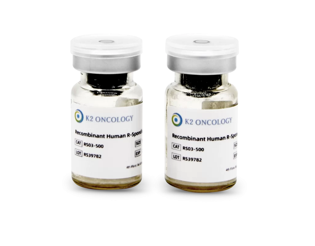
Recombinant Human R-Spondin 3 Protein
RS03-100
-

Recombinant Human R-Spondin 3 Protein
RS03-1000


Recombinant Human R-Spondin 3 Protein
RS03-100 包含以下产品
- Recombinant Human R-Spondin 3 Protein 冻干粉 100µg/vial x 1
RS03-1000 包含以下产品
- Recombinant Human R-Spondin 3 Protein 冻干粉 500µg/vial x 2

我们的科学家向您推荐
我们的产品简化了实验流程,集成多种因子,无需单独优化,扩增潜力高,14天内细胞数量可达到1×10^6。适用于多种培养形式,包括基质胶、低吸附孔板和生物反应器悬浮培养。GMP级别生产条件下制备,批次质量稳定,试剂含量是常规市售干细胞培养基的2倍,实现极佳的成本效益比。让复杂的培养变得简单快速,让科研变得更高效。
概览
产品参数:
Source: |
Human HEK293 cell line, HEK293-derived human R-Spondin 3 protein. | ||||||
Accession: | |||||||
Purity: | >90%, by SDS-PAGE under reducing conditions. | ||||||
Endotoxin Level: | <0.10 EU/μg of the protein by the LAL method. | ||||||
Activity: | Measured by its ability to induce Topflash reporter activity in HEK293 reporter cells. The ED50 for this effect is 0.5-5.0ng/mL in the presence of 20 ng/mL Recombinant Wnt Surrogate Fc Chimera Protein (K2 Oncology, Catalog # WT01-100). | ||||||
Structure: | Monomer | ||||||
Predicted Molecular Weight | 28.3 kDa (monomer). | ||||||
SDS-PAGE | 36-48 kDa, reducing conditions. | ||||||
Sterile: | 0.22μm sterile filtration. | ||||||
Product Form: | Lyophilized powder. | ||||||
Shipping & Storage: | The product is shipped at ambient temperature. Upon receipt, store it immediately at the temperature recommended below: Ø To the date of expiration, -20°C to -80°C as supplied. Ø 3 months, -20°C to -80°C under sterile conditions after reconstitution. Ø 1 month, 2 to 8 °C under sterile conditions after reconstitution. Avoid repeated freeze-thaw cycles. | ||||||
产品数据:


产品背景:
R-Spondin 3 (RSPO3), also known as Cristin 1 or roof plate-specific spondin 3, is a secreted protein with a molecular weight of approximately 36 kDa. It shares around 40% amino acid identity with other members of the R-Spondin family. All R-Spondins act as positive modulators of Wnt/β-catenin signaling, but each exhibits a distinct expression pattern. RSPO3, like other R-Spondins, contains two adjacent cysteine-rich furin-like domains (amino acids 35-135), a potential N-glycosylation site (amino acid 36), a thrombospondin (TSP-1) motif (amino acids 147-207), and a region rich in basic residues (amino acids 211-269). The furin-like domains are sufficient for stabilizing β-catenin. RSPO3 shares high amino acid sequence identity within amino acids 21-209 with mouse, rat, equine, bovine, and canine RSPO3 (93%, 92%, 97%, 96%, and 92% identity, respectively).
In mice, RSPO3 is crucial for the development of the placental labyrinthine layer, likely by promoting vascular development through VEGF expression . It is also essential for the expression of Gcm1, a placenta-specific transcription factor. RSPO3 is often expressed by or located near endothelial cells in mouse embryos. It is found in various regions, including the roof plate, tail, somites, otic vesicles, cephalic mesoderm, truncus arteriosus, atrioventricular canal of the developing heart, and developing limbs.
R-Spondins regulate Wnt/β-catenin signaling by competing with the Wnt antagonist DKK-1 for binding to the Wnt co-receptors LRP-6 and Kremen, thereby reducing DKK-1-mediated internalization.
| 产品名称 | 货号 | 规格 | 储存温度 | 保质期 |
|---|---|---|---|---|
| Recombinant Human R-Spondin 3 Protein | RS03-100/1000 | 100µg / 1mg | -20°C / -80°C | 3个月 |
类型
细胞因子
物种
人类
应用
细胞培养 / 类器官培养
商标
OrganoPro™
产品使用说明及支持信息
在产品文档中查找支持信息和使用说明,或在下方探索更多
| 文档类型 | 产品名称 | Catalog # |
|---|---|---|
| User manual | Recombinant Human R-Spondin 3 Protein | RS03-100 RS03-1000 |
资源及文献引用
相关资源及文献引用
Organoid drug screening report for a non-small cell lung cancer patient with EGFR gene mutation negativity: A case report and review of the literature
Pan, Yuetian, Hongshang Cui, and Yongbin Song | Frontiers in Oncology (2023)
Abstract:
Identification of solamargine as a cisplatin sensitizer through phenotypical screening in cisplatin-resistant NSCLC organoids
Han, Yi,et al. | Frontiers in Pharmacology (2022)
Abstract:
An Artemisinin Derivative ART1 Induces Ferroptosis by Targeting the HSD17B4 Protein Essential for Lipid Metabolism and Directly Inducing Lipid Peroxidation.
Xie, Jingjing, et al. | CCS Chemistry (2022)
Abstract:
Artemisinin and its derivatives, commonly known as antimalarial drugs, have gradually come to be regarded as potential antitumor agents, although their cytotoxic efficacy and mechanisms of action remain to be settled. Herein, we report that an artemisinin analog, ART1, can potently induce ferroptosis in a subset of cancer cell lines. Structure–activity relationship (SAR) analysis reveals that both the endoperoxide moiety and the artemisinin skeleton are required for the antitumor activity of ART1. Aided with ART1-based small-molecule tools, chemical proteomic analysis identified the HSD17B4 protein as a direct target of ART1. HSD17B4 resides in peroxisomes and is an essential enzyme in the catabolism of very-long-chain fatty acids. Our results demonstrate that ART1 initiates ferroptosis through selective oxidation of the fatty acids in peroxisomes by hijacking the HSD17B4 protein without disturbing its enzymatic function, providing a promising mechanism to develop therapeutics for cancer treatment. Read More: https://doi.org/10.31635/ccschem.021.202000691Pyrotinib in patients with HER2-amplified advanced non–small cell lung cancer: A prospective, multicenter, single-arm trial
Song, Zhengbo,et al. | Clinical Cancer Research (2022)
Abstract:
Glutamine synthetase licenses APC/C-mediated mitotic progression to drive cell growth
Zhao, Jiang-Sha,et al. | Nature Metabolism (2022)
Abstract:
Halofuginone sensitizes lung cancer organoids to cisplatin via suppressing PI3K/AKT and MAPK signaling pathways
Li, Hefei,et al. | Frontiers in Cell and Developmental Biology (2021)
Abstract:
Lung cancer is the leading cause of cancer death worldwide. Cisplatin is the major DNA-damaging anticancer drug that cross-links the DNA in cancer cells, but many patients inevitably develop resistance with treatment. Identification of a cisplatin sensitizer might postpone or even reverse the development of cisplatin resistance. Halofuginone (HF), a natural small molecule isolated from Dichroa febrifuga, has been found to play an antitumor role. In this study, we found that HF inhibited the proliferation, induced G0/G1 phase arrest, and promoted apoptosis in lung cancer cells in a dose-dependent manner. To explore the underlying mechanism of this antitumor effect of halofuginone, we performed RNA sequencing to profile transcriptomes of NSCLC cells treated with or without halofuginone. Gene expression profiling and KEGG analysis indicated that PI3K/AKT and MAPK signaling pathways were top-ranked pathways affected by halofuginone. Moreover, combination of cisplatin and HF revealed that HF could sensitize the cisplatin-resistant patient-derived lung cancer organoids and lung cancer cells to cisplatin treatment. Taken together, this study identified HF as a cisplatin sensitizer and a dual pathway inhibitor, which might provide a new strategy to improve prognosis of patients with cisplatin-resistant lung cancer.
COA查询
根据货号和批次号,在线查询已购买产品的COA证书
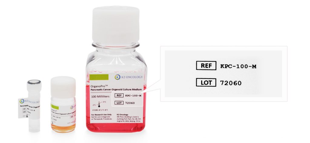
产品货号和产品批号均显示在产品标签上对应位置(如右侧示意图所示)

Ref/Cat#
产品货号,每个产品对应的独立货号
Lot#
批次编号,同一产品不同批次会有不同批号,请您在产品包装找到对应批号


